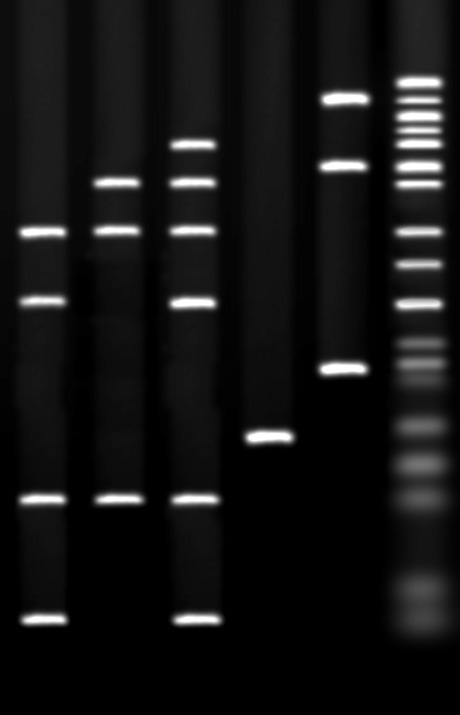
Ethidium Bromide Staining

The most commonly used stain for detecting DNA/RNA is ethidium bromide. Ethidium bromide is a DNA interchelator, inserting itself into the spaces between the base pairs of the double helix. Ethidium bromide possesses UV absorbance maxima at 300 and 360 nm. Additionally, it can absorb energy from nucleotides excited by the absorbance of 260 nm radiation. Ethidium re-emits this energy as yellow/orange light centered at 590 nm. The fluorescence of ethidium bromide in an aqueous solution is significantly lower than that of the interchelated dye.
Ethidium bromide is a sensitive, easy stain for DNA. It yields low background and a detection limit of 1-5 ng /band. The major drawback to ethidium bromide is that it is a potent mutagen. Solutions must be handled with extreme caution and decontaminated prior to disposal. Nonetheless, the sensitivity, simplicity ( the dye may be run in the gel with the DNA if desired, eliminating a separate staining/destaining process), and nondestructive nature of ethidium bromide staining have made it the standard stain for double-stranded DNA.
It is important to note that ethidium staining is strongly enhanced by the double stranded structure of native DNA. Staining of denatured, ssDNA or RNA is relatively insensitive, requiring some 10 fold more nucleic acid for equivalent detection. Another limitation is that the fluorescence of ethidium is quenched by polyacrylamide, reducing sensitivity by 10-20 fold in PAGE gels.
Detection of DNA/RNA using Ethidium Bromide
CAUTION: ETHIDIUM BROMIDE IS A POTENT MUTAGEN. HANDLE ONLY WITH GLOVES AND PROPER PRECAUTIONS.
Method I - Including Ethidium Bromide in the Gel and Buffer
GEL PREPARATION
- Dissolve agarose in the buffer as per the standard protocol for preparing an agarose gel.
- Allow the gel to cool to 60-70°C.
- Add EtBr to 0.5 µg/ml final concentration. (Stocks are generally 10 mg/ml, and require 5µl stock/100ml gel).
- Pour gel and allow to set as usual.
BUFFER PREPARATION
RUN GEL
Upon completion of the run, place gel in plastic wrap on a UV lightbox. Bands will appear bright orange on a faint orange background.
This method will detect approximately 5ng of DNA. Destaining in water or 1 mM MgSO4 may be required to achieve full sensitivity.
As an alternative, ethidium may be included in the gel, but not the buffer. Ethidium is positively charged and will migrate in the opposite direction from the DNA. In general, sufficient ethidium will remain bound to the DNA even at the cathode end of the gel. Such gels will have an area of high background where the ethidium has not yet migrated out of the gel. They are, however, sufficient for many purposes, and do not generate as much ethidium waste (Your safety office will know what level of ethidium causes a buffer to be declared a hazard).
Method II - Post-Run Staining
- Prepare enough 0.5µg/ml EtBr in water or buffer to completely submerge the gel. This solution is stable for 1-2 months at room temperature in the dark.
- After the run submerge the gel in the staining solution for 15-30 minutes (depending upon gel thickness).
- Place the gel on plastic wrap on a UV lightbox and observe under 300nm illumination. Bands will appear bright orange on a pale orange background.
NOTES:
This protocol minimizes the amount of ethidium bromide waste created with each gel run.
Sensitivity is the same as Method I, and may require destaining in water or 1mM MgSO4 to achieve the best sensitivity.
In this method, bands become visible from the top and bottom of the gel as the dye diffuses into the matrix. High contrast results can often be achieved without destaining by soaking the gel until the top and bottom of the bands appear and then leaving the gel to stand out of the staining solution for 15-30 minutes. During this time the stain will continue to diffuse into the gel, binding to the DNA at the expense of free dye. The result is a lower background without destaining.
Always use plastic wrap under ethidium-stained gels to avoid solarization damage to the surface of the transilluminator.
NEXT TOPIC: Silver Staining DNA Gels
