Post Electrophoretic Analysis
Faint bands, low background

Faint bands over a low background with standard Coomassie Blue R-250 staining can be caused by a number of factors: (scroll down for Colloidal Coomassie troubleshooting)
SDS in the staining solution: Most users save and re-use their staining solutions. This is fine for a couple of cycles, but over time enough SDS elutes from the gels to accumulate to significant levels in the staining reagent. SDS interferes with the binding of the dye to the proteins, resulting in bands that are fainter than expected. There is no practical way to regenerate the staining solution- discard it and make a fresh batch.

ProtoGel is your pathway to higher resolution, low backgrounds, and clearer gels.
To prolong the life of your staining solution, wash the gels briefly in water, or pre-fix the gels in methanol:water: acetic acid prior to staining.
Old dye or staining solution: Coomassie Blue R-250 is not stable indefinitely- older batches of dye or staining solution may show enough dye degradation to limit staining. In some cases, the solution will show a dye precipitate, and staining can be improved by filtering the mixture.
Staining time too short, or too little agitation: It takes time for the dye to penetrate the gel- the speed of this diffusion is increased substantially by agitation during staining. In general, you should not expect your darkest bands to be darker than the background when you begin destaining- ie the protein bands won't grow darker as the dye is eluted from the gel.

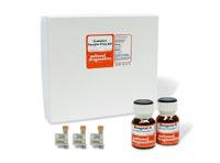
The ProtoGel Sample Prep Kit clears your sample for high-resolution results.
Bands too diffuse to see, due to poor electrophoretic separation: Always use fresh, high-quality matrix and buffer components- poor reagents will result in electroendosmotic band spreading, which can dramatically lower band intensities.
Too little protein-loaded: Coomassie Blue R-250 has a detection limit of approximately 100ng per band with Bovine Serum Albumin (BSA), but this limit varies widely among different proteins. Concentrate dilute samples to ensure loading enough to detect.

The ProtoGel Sample Prep Kit clears your sample for high resolution results.
ProtoBlue Safe/Colloidal Coomassie:

ProtoBlue Safe's fast, easy protocol increases the likelihood of clear backgrounds in your sample.
The most frequent cause of faint banding over the low background with ProtoBlue Safe is the failure to agitate the dye solution prior to decanting and adding ethanol. The dye in ProtoBlue Safe is in a colloidal form, so it settles out of the solution on standing. If it is not mixed back in before an aliquot is removed, the dye solution will not have enough Coomassie in it to sufficiently stain the proteins. This does no harm to the gel; staining with another batch of ProtoBlue will give the full intensity of detection.
