Post Electrophoretic Analysis
Blotches on Gel

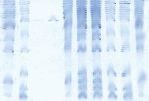
This gel can be recovered in most cases by fully destaining in methanol:water: acetic acid, followed by a fresh round of staining. In severe cases, the deposits may be removed by gently spraying the gel surface with alcohol.

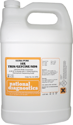
Blotches can be caused by overhandling of the gel, or by handling the gel without gloves. This can create uneven surfaces or protein deposits that can bind Coomassie to the gel. To minimize this problem, handle the gel as little as possible, and always with clean gloves – have us prepare a gallon of ready-made 10X Tris-Glycine-SDS Buffer for you!
Occasionally blotches result from using a dye solution that is not completely dissolved- The dye crystals deposit on the gel surface and produce an area of the intense background. This will eventually disappear during destaining.
- UV Shadowing
- Uneven Staining
- Staining Proteins Immobilized on Membranes
- Staining Protein Gels with Coomassie Blue
- Southern Blotting
- Smeared Bands
- Silver Staining Protein Gels
- Silver Staining DNA Gels
- Protein Fixation on Gels
- Post-Electrophoretic Visualization with Nuclistain
- Overview of Western Blotting
- Northern Blotting
- Method for Western Blotting
- Mechanism of Immunostaining
- Mechanism of Immunostaining
- Immunostaining with Alkaline Phosphatase
- Guide Strip Technique
- Faint bands, low background
- Faint Bands, High Background
- Ethidium Bromide Staining
- Enzyme Linked Immunosorbent Assay (ELISA)
- Coomassie Blue Stain- Troubleshooting
- Blotches on Gel
- Autoradiography
- Autoradiographic Enhancement with Autofluor
- An Overview of Northern and Southern Blotting
- Alkaline Blotting


