Electrophoresis
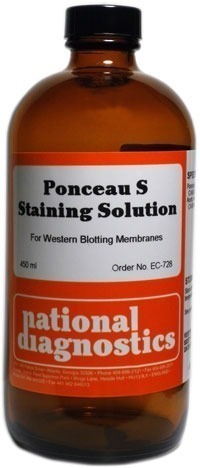
Ponceau S Staining Solution
$57.00
Catalog Number: EC-728
Size: 450 ml
Size: 450 ml
- Ideal for Western blotting membranes
- Premixed formula saves time, money, and effort
- Stain up to 90 mini blots
Description
Catalog Number: EC-728
Size: 450 ml
Size: 450 ml
- Ideal for Western blotting membranes
- Premixed formula saves time, money, and effort
- Stain up to 90 mini blots
National Diagnostics’ Ponceau S Staining Solution is optimal for staining proteins that have been transferred to PVDF or nitrocellulose membranes from polyacrylamide gels.
Additional information
Weight | 4.6 lbs |
---|
- UV Shadowing
- Using PAGE to Determine Nucleic Acid Molecular Weight
- Uneven Staining
- The Polyacrylamide Matrix-Buffer Strength
- The Polyacrylamide Matrix
- The Mechanical and Electrical Dynamics of Gel Electrophoresis — Electrophoresis System Dynamics
- The Mechanical and Electrical Dynamics of Gel Electrophoresis – Ohm’s Law
- The Mechanical and Electrical Dynamics of Gel Electrophoresis – Intro and Sample Mobility
- The Electrophoresis Matrix
- The Agarose Matrix
- Staining Proteins Immobilized on Membranes
- Staining Protein Gels with Coomassie Blue
- SSCP Analysis
- Southern Blotting
- Smeared Bands
- Silver Staining Protein Gels
- Silver Staining DNA Gels
- Sanger Sequencing
- Sample Preparation for SDS-PAGE
- Sample Preparation for Native Protein Electrophoresis
- Sample Preparation for Native PAGE of DNA
- Sample Prep for Denaturing PAGE of DNA
- S1 Mapping
- Run Conditions in Denaturing PAGE
- RNA Mapping
- RNA Electrophoresis
- Ribonuclease Protection
- Restriction Digest Mapping
- Radioactive Emissions and the Use of Isotopes in Research
- Protein Fixation on Gels
- Primer Extension
- Preparing Denaturing DNA & RNA Gels
- Preparation of Denaturing Agarose Gels
- Preparation of Agarose Gels
- Pouring Sequencing Gels
- Post-Electrophoretic Visualization with Nuclistain
- PFGE and FIGE
- Peptide Mapping
- PCR Analysis: Yield and Kinetics
- PCR Analysis: An Examination
- Overview of Western Blotting
- Northern Blotting
- Native Protein Electrophoresis
- Native PAGE of DNA
- Multiphasic Buffer Systems
- Mobility Shift Assay
- Methylation & Uracil Interference Assays
- Method for Western Blotting
- Mechanism of Immunostaining
- Measuring Molecular Weight with SDS-PAGE
- Maxam & Gilbert Sequencing
- Manual Sequencing
- Isotachophoresis
- Isoelectric Focusing
- In Gel Enzyme Reactions
- Immunostaining with Alkaline Phosphatase
- Immuno-Electrophoresis / Immuno-Diffusion
- Horizontal and Vertical Gel Systems – Vertical Tube Gels
- Horizontal and Vertical Gel Systems – The Vertical Slab Gel System
- Horizontal and Vertical Gel Systems – The Horizontal Gel System
- Homogeneous Buffer Systems
- Heteroduplex Analysis
- Guide Strip Technique
- Gel Preparation for Native Protein Electrophoresis
- Gel Preparation for Native PAGE of DNA
- Gel Electrophoresis of RNA & Post Electrophoretic Analysis
- Gel Electrophoresis of PCR Products
- Faint bands, low background
- Faint Bands, High Background
- Ethidium Bromide Staining
- Enzyme Linked Immunosorbent Assay (ELISA)
- Electrophoresis Buffers-Choosing the Right Buffer
- Electrophoresis Buffers–The Henderson-Hasselbalch Equation
- DNase I Footprinting
- DNA/RNA Purification from PAGE Gels
- DNA/RNA Purification from Agarose Gels – Electroelution
- Differential Display
- Denaturing Protein Electrophoresis: SDS-PAGE
- Denaturing Polyacrylamide Gel Electrophoresis of DNA & RNA
- Coomassie Blue Stain- Troubleshooting
- Conformational Analysis
- Casting Gradient Gels
- Buffer Additives-Surfactants
- Buffer Additives-Reducing Agents
- Buffer Additives-Hydrogen Bonding Agents
- Blotches on Gel
- Biological Macromolecules: Nucleic Acids
- Biological Macromolecules – Proteins
- Autoradiography
- Autoradiographic Enhancement with Autofluor
- Automated Sequencers
- Analysis of DNA/Protein Interactions
- An Overview of Northern and Southern Blotting
- Alkaline Blotting
- Agarose Gel Electrophoresis of DNA and RNA – Uses and Variations
- Agarose Gel Electrophoresis of DNA and RNA – An Introduction
- Activity Stains