Electrophoresis
ProtoMarkers
$147.00
Size: 0.5 ml
- Proteins Stained with High Definition Blue Dye
- Red Protein Band Included for Easy Orientation
- High Contrast, High-Intensity Labeling
Description
Size: 0.5 ml
- Proteins Stained with High Definition Blue Dye
- Red Protein Band Included for Easy Orientation
- High Contrast, High-Intensity Labeling
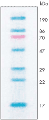
 ProtoMarkers consist of seven (7) purified proteins. Six markers are permanently labeled with high-contrast blue dye. One protein is labeled with high-contrast red dye to facilitate accurate positioning on the gel.
ProtoMarkers Protein Standards range in size from approximately 20 kD to 190 kD, covering the most common protein molecular weights. Marker sizes shown are representative- each lot of ProtoMarkers is individually calibrated to provide accurate size information for the labeled proteins in that lot.
ProtoMarkers are supplied in quantities of 500 microliters per vial. Each vial contains sufficient material for 100 mini-gels.
Additional information
| Weight | 4 lbs |
|---|---|
| Dimensions | 8 × 6 × 6 in |
Safety Overview
Safety Summary (see SDS for complete information before using product):
Appearance and Odor
Clear, colorless solution
EMERGENCY OVERVIEW – IMMEDIATE HAZARD
2-Mercaptoethanol
DANGER! MAY BE FATAL IF ABSORBED THROUGH SKIN. HARMFUL IF SWALLOWED OR INHALED. CAUSES IRRITATION TO SKIN, EYES, AND RESPIRATORY TRACT. COMBUSTIBLE LIQUID AND VAPOR. Health hazards given on this data sheet apply to concentrated solutions of mercaptoethanol or the substance in its pure form. Hazards of dilute solutions may be reduced, depending upon the concentration. Degree of hazard for these reduced concentrations is not currently addressed in the available literature.
Tris-Base
CAUSES IRRITATION TO SKIN, EYES, AND RESPIRATORY TRACT. HARMFUL IF SWALLOWED OR INHALED.
- UV Shadowing
- Using PAGE to Determine Nucleic Acid Molecular Weight
- Uneven Staining
- The Polyacrylamide Matrix-Buffer Strength
- The Polyacrylamide Matrix
- The Mechanical and Electrical Dynamics of Gel Electrophoresis — Electrophoresis System Dynamics
- The Mechanical and Electrical Dynamics of Gel Electrophoresis – Ohm’s Law
- The Mechanical and Electrical Dynamics of Gel Electrophoresis – Intro and Sample Mobility
- The Electrophoresis Matrix
- The Agarose Matrix
- Staining Proteins Immobilized on Membranes
- Staining Protein Gels with Coomassie Blue
- SSCP Analysis
- Southern Blotting
- Smeared Bands
- Silver Staining Protein Gels
- Silver Staining DNA Gels
- Sanger Sequencing
- Sample Preparation for SDS-PAGE
- Sample Preparation for Native Protein Electrophoresis
- Sample Preparation for Native PAGE of DNA
- Sample Prep for Denaturing PAGE of DNA
- S1 Mapping
- Run Conditions in Denaturing PAGE
- RNA Mapping
- RNA Electrophoresis
- Ribonuclease Protection
- Restriction Digest Mapping
- Radioactive Emissions and the Use of Isotopes in Research
- Protein Fixation on Gels
- Primer Extension
- Preparing Denaturing DNA & RNA Gels
- Preparation of Denaturing Agarose Gels
- Preparation of Agarose Gels
- Pouring Sequencing Gels
- Post-Electrophoretic Visualization with Nuclistain
- PFGE and FIGE
- Peptide Mapping
- PCR Analysis: Yield and Kinetics
- PCR Analysis: An Examination
- Overview of Western Blotting
- Northern Blotting
- Native Protein Electrophoresis
- Native PAGE of DNA
- Multiphasic Buffer Systems
- Mobility Shift Assay
- Methylation & Uracil Interference Assays
- Method for Western Blotting
- Mechanism of Immunostaining
- Mechanism of Immunostaining
- Measuring Molecular Weight with SDS-PAGE
- Maxam & Gilbert Sequencing
- Manual Sequencing
- Isotachophoresis
- Isoelectric Focusing
- In Gel Enzyme Reactions
- Immunostaining with Alkaline Phosphatase
- Immuno-Electrophoresis / Immuno-Diffusion
- Horizontal and Vertical Gel Systems – Vertical Tube Gels
- Horizontal and Vertical Gel Systems – The Vertical Slab Gel System
- Horizontal and Vertical Gel Systems – The Horizontal Gel System
- Homogeneous Buffer Systems
- Heteroduplex Analysis
- Guide Strip Technique
- Gel Preparation for Native Protein Electrophoresis
- Gel Preparation for Native PAGE of DNA
- Gel Electrophoresis of RNA & Post Electrophoretic Analysis
- Gel Electrophoresis of PCR Products
- Faint bands, low background
- Faint Bands, High Background
- Ethidium Bromide Staining
- Enzyme Linked Immunosorbent Assay (ELISA)
- Electrophoresis Buffers-Choosing the Right Buffer
- Electrophoresis Buffers–The Henderson-Hasselbalch Equation
- DNase I Footprinting
- DNA/RNA Purification from PAGE Gels
- DNA/RNA Purification from Agarose Gels – Electroelution
- Differential Display
- Denaturing Protein Electrophoresis: SDS-PAGE
- Denaturing Polyacrylamide Gel Electrophoresis of DNA & RNA
- Coomassie Blue Stain- Troubleshooting
- Conformational Analysis
- Casting Gradient Gels
- Buffer Additives-Surfactants
- Buffer Additives-Reducing Agents
- Buffer Additives-Hydrogen Bonding Agents
- Blotches on Gel
- Biological Macromolecules: Nucleic Acids
- Biological Macromolecules – Proteins
- Autoradiography
- Autoradiographic Enhancement with Autofluor
- Automated Sequencers
- Analysis of DNA/Protein Interactions
- An Overview of Northern and Southern Blotting
- Alkaline Blotting
- Agarose Gel Electrophoresis of DNA and RNA – Uses and Variations
- Agarose Gel Electrophoresis of DNA and RNA – An Introduction
- Activity Stains


