Electrophoresis Articles
Smeared Bands
Band distortions are not staining artifacts, they occur during the electrophoretic run. Some of the most common causes are listed below: Particulate in the sample: Standard loading buffers are often unable to completely dissolve complex samples- fragments of bacterial cell walls, for example, can be carried over into the well. Particulate material in the well…
Read MoreBlotches on Gel
Blotches can be caused by overhandling of the gel, or by handling the gel without gloves. This can create uneven surfaces or protein deposits that can bind Coomassie to the gel. To minimize this problem, handle the gel as little as possible, and always with clean gloves. This gel can be recovered in most…
Read MoreUneven Staining
Uneven staining is almost always the result of insufficient agitation during staining. The gel is initially more dense than the staining solution and will tend to sink to the bottom of the dish. Some portions of the gel will have stains under them, others will not, and the gel will show darker and lighter areas…
Read MoreFaint Bands, High Background
A high background which obscures the bands, or in combination with fainter than usual bands, indicates dye binding to the gel matrix, or contamination of the matrix with a dye-binding material (most often a protein). Coomassie Blue R-250: Destaining time too short: It takes hours for the dye to completely diffuse out of the gel…
Read MoreFaint bands, low background
Faint bands over a low background with standard Coomassie Blue R-250 staining can be caused by a number of factors: (scroll down for Colloidal Coomassie troubleshooting) SDS in the staining solution: Most users save and re-use their staining solutions. This is fine for a couple of cycles, but over time enough SDS elutes from the…
Read MoreCoomassie Blue Stain- Troubleshooting
If your gel doesn’t look like this one, click on the problem below to find the solution: Faint bands on a low background Learn More Faint bands on a high background Learn More Uneven Staining Learn More Dark Blotches on Gel Learn More Smeared or blurred bands Learn More The ProtoGel Sample Prep Kit gives…
Read MoreMechanism of Immunostaining
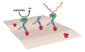
The basic method of immunostaining is to probe with an antibody that is bound to a detectable molecule such as an enzyme or a fluorescent dye. The highly specific binding interaction between antibodies and their unique antigens has been exploited to create sensitive and specific detection systems for proteins. An antibody can be raised and/or…
Read MoreDNase I Footprinting
A method for determining the location of a protein binding site, DNase I Footprinting Analysis involves endonuclease treatment of an end labeled DNA fragment bound to a protein. Limited digestion yields fragments terminating everywhere except in the footprint region, which is protected from digestion. DNase I Footprinting Procedure Preparing the DNA substrate: DNA to be…
Read MoreRadioactive Emissions and the Use of Isotopes in Research
Radioactive decay occurs with the emission of particles or electromagnetic radiation from an atom due to a change within its nucleus. Forms of radioactive emission include alpha particles (α), beta particles (β), and gamma rays (γ). α particles are the least energetic, most massive of these decay products. An α particle contains two protons and…
Read MoreMethod for Western Blotting
ELECTROPHORESIS Prepare and run an SDS PAGE gel. Select a gel percent which will give the best resolution for the size of antigen being analyzed (if known). If the size is not known, a 12% gel is a good starting point. Load enough protein to provide 0.1-1ng of antigen per well. It is generally advantageous…
Read More


