Post Electrophoretic Analysis
Mechanism of Immunostaining

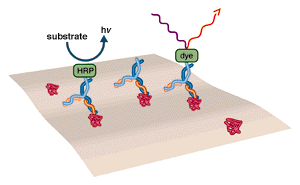
The basic method of immunostaining is to probe with an antibody that is bound to a detectable molecule such as an enzyme or a fluorescent dye.
The highly specific binding interaction between antibodies and their unique antigens has been exploited to create sensitive and specific detection systems for proteins. An antibody can be raised and/or purified “against” (i.e. binding to) a single “epitope” (area bound by the antibody) on a larger “antigen” (a molecule containing many epitopes). When antibody and antigen are mixed, the antibody will bind tightly to the epitope it recognizes. This identifies the antigen as bearing the epitope in question. Thus, immunological detection answers questions about the structure and identity of a protein which cannot be addressed through conventional chemical stains. In addition, immunological detection can be as much as 100-fold more sensitive than chemical stains.
Antibodies, or immunoglobulins, are produced by the body in response to invasion by foreign proteins. The function of an antibody is to bind to a specific site (epitope) on the invader, thus tagging it for destruction by other agents of the immune system. The salient characteristics of antibody binding are binding strength and selectivity. In assay terminology, antibodies are said to be accurate (low false negatives) and specific (low false positives).
In the body, the mere presence of a bound antibody on the surface of an antigen is a sufficient signal for the immune system to “detect” that molecule. In an in-vitro assay, additional aids are required. These aids take the form of easily detectable molecules, which are bound to the antibody. Examples are enzymes, such as horseradish peroxidase (HRP) or fluorescent dyes. Enzymes are particularly suited to this role, because they are proteins, and thus stable wherever antibodies are stable. The catalytic cycling of enzymes greatly multiplies the sensitivity of the assay. The two enzymes most commonly used for labeling antibodies are horseradish peroxidase and alkaline phosphatase (AP). Chromogenic and luminogenic substrates are available for both of these enzymes.
NEXT TOPIC: Immunostaining with Alkaline Phosphatase
- UV Shadowing
- Uneven Staining
- Staining Proteins Immobilized on Membranes
- Staining Protein Gels with Coomassie Blue
- Southern Blotting
- Smeared Bands
- Silver Staining Protein Gels
- Silver Staining DNA Gels
- Protein Fixation on Gels
- Post-Electrophoretic Visualization with Nuclistain
- Overview of Western Blotting
- Northern Blotting
- Method for Western Blotting
- Mechanism of Immunostaining
- Mechanism of Immunostaining
- Immunostaining with Alkaline Phosphatase
- Guide Strip Technique
- Faint bands, low background
- Faint Bands, High Background
- Ethidium Bromide Staining
- Enzyme Linked Immunosorbent Assay (ELISA)
- Coomassie Blue Stain- Troubleshooting
- Blotches on Gel
- Autoradiography
- Autoradiographic Enhancement with Autofluor
- An Overview of Northern and Southern Blotting
- Alkaline Blotting
