Electrophoresis
Insite Markers
$147.00
Catalog number: EC-897
Size: 0.5 ml
Size: 0.5 ml
- Two Sets of Markers in One
 - Prestained Markers Provide Orientation During the Run
 - Engineered Molecular Weight Standards Appear in Fluorescent Detection
 
Description
Catalog number: EC-897
Size: 0.5 ml
Size: 0.5 ml
- Two Sets of Markers in One
 - Prestained Markers Provide Orientation During the Run
 - Engineered Molecular Weight Standards Appear in Fluorescent Detection
 

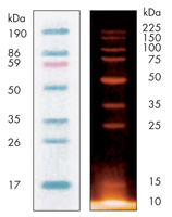
National Diagnostics’ Insite Markers contain both visible markers for orientation during the run and high precision protein standards (10-225 kD) that appear with fluorescent detection. This allows both confident monitoring of the run and precise assignment of protein molecular weights.
The Insite Markers are supplied in a 0.5 mL vial. Each vial contains sufficient material to stain between 50 and 100 mini-gels.
Additional information
| Weight | 4 lbs | 
|---|---|
| Dimensions | 8 × 6 × 6 in | 
Protocol
Twenty Minute Casting
ProtoGel Quick-Cast contains the monomers and buffer components to produce a 12% gel.
- Measure out the volume of ProtoGel Quick-Cast needed to fill the cassette – typically 10ml for one mini-gel, 15ml for two.
 - Add 100 microliters of fresh 10% APS and 10 microliters of TEMED per 10ml ProtoGel Quick-Cast. Mix briefly and pour into the gel cassette.
 - 3. Insert comb and allow to polymerize at room temperature for 20 minutes. The gel is now ready to run.
 
For best results, it is recommended you use ProtoGel Quick-Cast Loading Buffer. Simply mix your samples with an equal volume of ProtoGel Quick-Cast Loading Buffer, load and run.
- UV Shadowing
 - Using PAGE to Determine Nucleic Acid Molecular Weight
 - Uneven Staining
 - The Polyacrylamide Matrix-Buffer Strength
 - The Polyacrylamide Matrix
 - The Mechanical and Electrical Dynamics of Gel Electrophoresis — Electrophoresis System Dynamics
 - The Mechanical and Electrical Dynamics of Gel Electrophoresis – Ohm’s Law
 - The Mechanical and Electrical Dynamics of Gel Electrophoresis – Intro and Sample Mobility
 - The Electrophoresis Matrix
 - The Agarose Matrix
 - Staining Proteins Immobilized on Membranes
 - Staining Protein Gels with Coomassie Blue
 - SSCP Analysis
 - Southern Blotting
 - Smeared Bands
 - Silver Staining Protein Gels
 - Silver Staining DNA Gels
 - Sanger Sequencing
 - Sample Preparation for SDS-PAGE
 - Sample Preparation for Native Protein Electrophoresis
 - Sample Preparation for Native PAGE of DNA
 - Sample Prep for Denaturing PAGE of DNA
 - S1 Mapping
 - Run Conditions in Denaturing PAGE
 - RNA Mapping
 - RNA Electrophoresis
 - Ribonuclease Protection
 - Restriction Digest Mapping
 - Radioactive Emissions and the Use of Isotopes in Research
 - Protein Fixation on Gels
 - Primer Extension
 - Preparing Denaturing DNA & RNA Gels
 - Preparation of Denaturing Agarose Gels
 - Preparation of Agarose Gels
 - Pouring Sequencing Gels
 - Post-Electrophoretic Visualization with Nuclistain
 - PFGE and FIGE
 - Peptide Mapping
 - PCR Analysis: Yield and Kinetics
 - PCR Analysis: An Examination
 - Overview of Western Blotting
 - Northern Blotting
 - Native Protein Electrophoresis
 - Native PAGE of DNA
 - Multiphasic Buffer Systems
 - Mobility Shift Assay
 - Methylation & Uracil Interference Assays
 - Method for Western Blotting
 - Mechanism of Immunostaining
 - Mechanism of Immunostaining
 - Measuring Molecular Weight with SDS-PAGE
 - Maxam & Gilbert Sequencing
 - Manual Sequencing
 - Isotachophoresis
 - Isoelectric Focusing
 - In Gel Enzyme Reactions
 - Immunostaining with Alkaline Phosphatase
 - Immuno-Electrophoresis / Immuno-Diffusion
 - Horizontal and Vertical Gel Systems – Vertical Tube Gels
 - Horizontal and Vertical Gel Systems – The Vertical Slab Gel System
 - Horizontal and Vertical Gel Systems – The Horizontal Gel System
 - Homogeneous Buffer Systems
 - Heteroduplex Analysis
 - Guide Strip Technique
 - Gel Preparation for Native Protein Electrophoresis
 - Gel Preparation for Native PAGE of DNA
 - Gel Electrophoresis of RNA & Post Electrophoretic Analysis
 - Gel Electrophoresis of PCR Products
 - Faint bands, low background
 - Faint Bands, High Background
 - Ethidium Bromide Staining
 - Enzyme Linked Immunosorbent Assay (ELISA)
 - Electrophoresis Buffers-Choosing the Right Buffer
 - Electrophoresis Buffers–The Henderson-Hasselbalch Equation
 - DNase I Footprinting
 - DNA/RNA Purification from PAGE Gels
 - DNA/RNA Purification from Agarose Gels – Electroelution
 - Differential Display
 - Denaturing Protein Electrophoresis: SDS-PAGE
 - Denaturing Polyacrylamide Gel Electrophoresis of DNA & RNA
 - Coomassie Blue Stain- Troubleshooting
 - Conformational Analysis
 - Casting Gradient Gels
 - Buffer Additives-Surfactants
 - Buffer Additives-Reducing Agents
 - Buffer Additives-Hydrogen Bonding Agents
 - Blotches on Gel
 - Biological Macromolecules: Nucleic Acids
 - Biological Macromolecules – Proteins
 - Autoradiography
 - Autoradiographic Enhancement with Autofluor
 - Automated Sequencers
 - Analysis of DNA/Protein Interactions
 - An Overview of Northern and Southern Blotting
 - Alkaline Blotting
 - Agarose Gel Electrophoresis of DNA and RNA – Uses and Variations
 - Agarose Gel Electrophoresis of DNA and RNA – An Introduction
 - Activity Stains


