
Histology Fundamentals
Mounting Tissue Sections
To preserve and support a stained section for light microscopy, it is mounted on a clear glass slide, and covered with a thin glass coverslip. The slide and coverslip must be free of optical distortions, to avoid viewing artifacts. A mounting medium is used to adhere the coverslip to the slide. Aqueous based mounting media are available, which allow the mounting of tissues directly from the staining procedure. However, the water solubility of some stains allows them to bleed and/or fade in such mountants, necessitating the use of resinous mounting media. To use a nonaqueous mountant, the section must first be dehydrated (again!) and cleared. Any water carried over to the mounting stage will show up as bubbles or vacuole-like structures, as the water droplets aggregate and distort the tissue. It is important to note also that the clearing agent used must be compatible with the mounting medium, or the sections must be thoroughly dried prior to mounting.
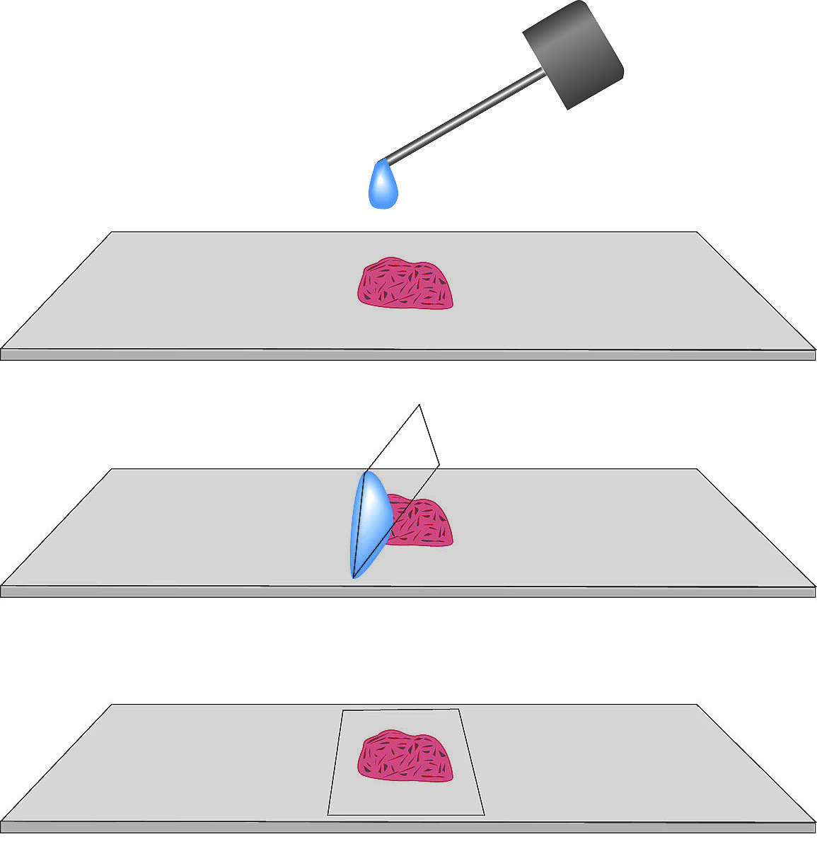
To mount a slide, (A) Apply a single drop of mounting medium upon tissue section. (B) Hold coverslip at 45º allowing the drop to spread along the edge of the slip. (C) Let go of slip and allow medium to spread slowly.
Mounting Slides with Histo-Clear and Histomount
- Drain excess Histo-Clear from the slide by standing on end on a paper towel. Wipe excess Histo-Clear from the back of the slide.
- Place the slide on a level surface, and apply a drop of Histomount using the dispenser rod.
- Hold the cover slip at a 45° angle to the surface of the slide, and allow the bottom edge to touch the drop of Histomount. When the drop has spread along the edge of the slip, let go of the slip and allow the Histomount to spread slowly (20-30 seconds).
- Excess mounting medium may be removed while wet with a tissue or with a razor blade when dry.Histomount will dry sufficiently to be read in 30 minutes. Full drying may require up to 48 hours. Drying can be accelerated at 37°C.
NEXT TOPIC: Immunohistochemistry
- Working Safely with Fixatives
- The Chemistry of Dyes and Staining
- Suggested procedures for processing fixed tissue
- Staining Procedures
- Sectioning
- Overview of the Paraffin Technique
- Overview of Fixation
- Non-Aldehyde Fixatives
- Mounting Tissue Sections
- Factors Affecting Fixation
- Embedding
- Dehydration
- Decalcifying Tissue for Histological Processing
- Clearing Tissue Sections
- Artifacts in Histologic Sections
- Aldehyde Fixatives