Post Electrophoretic Analysis
UV Shadowing

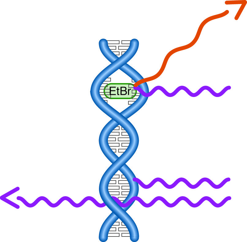
When ultraviolet light strikes a DNA molecule, it may be absorbed, transmitted, or, if a fluorescent dye is present, re-emitted as visible light.
Detection of DNA/RNA in solution is generally done by measuring its UV absorbance at 260 nm. This absorbance is due to the ring systems in the nitrogen bases, and can also be used for low sensitivity detection of DNA/RNA in gels. In a technique known as UV shadowing, the gel is placed on plastic wrap over a UV fluorescent TLC plate. The dye in the plate is excited by placing a near-UV source over the gel. Dark areas are observed where DNA in the gel absorbs the UV light. The sensitivity of this method is limited, on the order of 10-50 ng, and its use is limited to thin gels, which do not excessively attenuate the UV light.
NEXT TOPIC: Post-Electrophoretic Visualization with Nuclistain
- UV Shadowing
- Uneven Staining
- Staining Proteins Immobilized on Membranes
- Staining Protein Gels with Coomassie Blue
- Southern Blotting
- Smeared Bands
- Silver Staining Protein Gels
- Silver Staining DNA Gels
- Protein Fixation on Gels
- Post-Electrophoretic Visualization with Nuclistain
- Overview of Western Blotting
- Northern Blotting
- Method for Western Blotting
- Mechanism of Immunostaining
- Mechanism of Immunostaining
- Immunostaining with Alkaline Phosphatase
- Guide Strip Technique
- Faint bands, low background
- Faint Bands, High Background
- Ethidium Bromide Staining
- Enzyme Linked Immunosorbent Assay (ELISA)
- Coomassie Blue Stain- Troubleshooting
- Blotches on Gel
- Autoradiography
- Autoradiographic Enhancement with Autofluor
- An Overview of Northern and Southern Blotting
- Alkaline Blotting
