Advanced Histological Techniques
Detection Systems in Immunohistochemistry
Light microscopy makes use of primarily two detection systems for immunohistochemistry - fluorescence and enzyme labeling, while electron microscopy relies on the deposition of electron dense materials at the site of antibody binding. Techniques for light microscopy are discussed below. EM is covered briefly in the next section.
Immunoflourescence
The conjugation of a fluorescent dye to the primary or secondary antibody allows its detection under ultraviolet light. Microscopes are available which allow a slide to be UV illuminated, producing brightly emitting areas where the antibody has bound. Typically, Texas Red, rhodamine, fluorescine, or phycoerythrin are conjugated to the antibody. Texas Red and rhodamine fluoresce in the red range, fluorescine in the green and phycoerythrin in the blue. This allows multiple label experiments, in which different antigens show up as different colors. While extremely sensitive, this technique suffers from the fact that the dyes fade with time. Long term storage of slides is seldom practical, as loss of signal destroys their utility.
Enzyme-Antibody Conjugate Methods
Methods have been developed to detect some enzymes at extremely low levels. These enzymes have been used as labels to facilitate detection of various molecules. Horseradish peroxidase (HRP) is often conjugated to antibodies, producing a system capable of detecting antigens by peroxidase staining. Incubation of HRP in a solution containing hydrogen peroxide and diaminobenzidine (DAB) results in the reduction of DAB to an insoluble brown precipitate, visible under the light microscope. Alkaline phosphatase (AP) is also used in an analogous method, in which case a phosphorylated naphthol is used as the substrate. The dephosphorylated napthol reacts with an azo dye such as Fast Red TR, to give a colored reaction product.
A level of signal amplification is provided by the peroxidase-antiperoxidase complex system (PAP). This technique uses large complexes which contain many detection agents bound together. The binding of one of these complexes to each secondary antibody amplifies the signal manyfold.
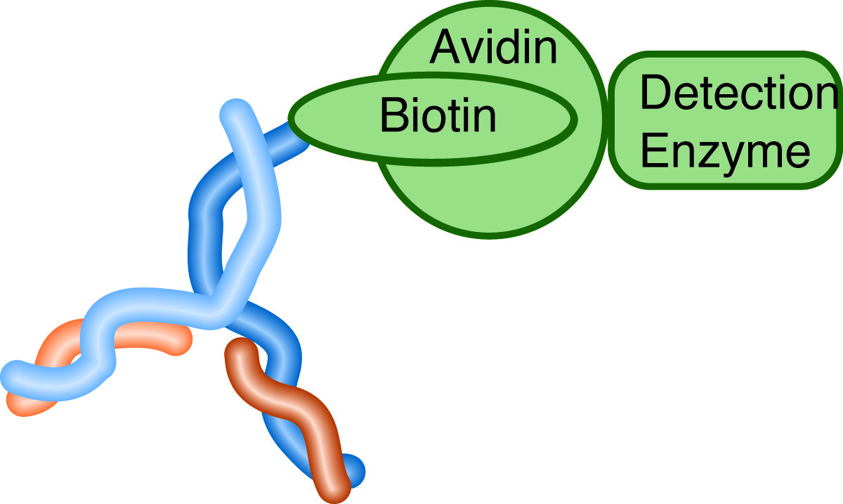
Either of these techniques may be combined with the Avidin-Biotin system to provide a more robust and easily generalizable detection method. Biotin is a small molecule which is bound tightly and specifically by the protein avidin. This binding has been exploited by attaching biotin to the secondary antibody (biotinylation), and avidin to the detection enzyme. Some of the advantages of this approach are:
- Only a small molecule is attached to the antibody, which minimizes the chance of interfering with the antibody-antigen interaction.
- The same secondary antibody may be used with many detection molecules, simply by attaching avidin to each.
- Use of an avidin-biotin complex as a bridging reagent amplifies the signal, as many detection molecules will bind to each biotinylated antibody. This is the same amplification mechanism outlined for the PAP system above.
 The use of biotinylated antibodies and avidin attached detection enzymes increases sensitivity, flexibility and reliability
NEXT TOPIC: Electron Microscopy
