Histology
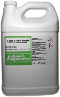
Calci-Clear Rapid
$36.00 – $455.00
Catalog number: HS-105
- High-speed decalcifier
- For urgent specimens
Description
Catalog number: HS-105
- High-speed decalcifier
- For urgent specimens
Additional information
| Weight | 3 lbs |
|---|---|
| Dimensions | 15 × 10 × 13 in |
Protocol
Procedure
- Rinse fixed tissue in running water for five minutes.
- Submerge the tissue sample in a volume of Calci-Clear Rapid equal to at least ten times the volume of the sample. Take care that the Calci-Clear Rapid has free access to all surfaces of the tissue.
- Check tissue every thirty to sixty minutes. Although some workers consider decalcification complete when the tissue begins to float, for some types of tissue this is not an accurate indicator of the endpoint of decalcification. A chemical test may be employed for to more accurately confirm the endpoint of decalcification.
- Changing the solution is recommended every two or three hours.
Safety Overview
Safety Summary (see SDS for complete information before using product):
Appearance and Odor
Clear liquid.
EMERGENCY OVERVIEW – IMMEDIATE HAZARD
DANGER! CORROSIVE. LIQUID AND MIST CAUSE SEVERE BURNS TO ALL BODY TISSUE. MAY BE FATAL IF SWALLOWED OR INHALED. Health hazards given on this data sheet apply to concentrated solutions of hydrochloric acid. Hazards of dilute solutions may be reduced, depending upon the concentration. Degree of hazard for these reduced concentrations is not currently addressed in the available literature.
Causes Burns Keep locked up and out of reach of children. Do not breathe fumes. In case of contact with eyes, rinse immediately with plenty of water and seek medical advice. In case of accident or if you feel unwell, seek medical advice immediately (show label where possible).
- Working Safely with Fixatives
- Tissue Processing for Electron Microscopy
- The Chemistry of Dyes and Staining
- Suggested procedures for processing fixed tissue
- Staining Tissue Sections for Electron Microscopy
- Staining Procedures
- Sectioning Tissue for Electron Microscopy
- Sectioning
- Overview of the Paraffin Technique
- Overview of Fixation
- Non-Aldehyde Fixatives
- Mounting Tissue Sections
- Immunohistochemistry
- Fixing Tissue for Electron Microscopy
- Factors Affecting Fixation
- Embedding
- Electron Microscopy
- Detection Systems in Immunohistochemistry
- Dehydration
- Decalcifying Tissue for Histological Processing
- Clearing Tissue Sections
- Artifacts in Histologic Sections
- Antibody Binding
- Aldehyde Fixatives


