Electrophoresis
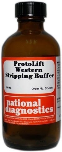
ProtoLift Western Stripping Buffer
$47.00
Catalog number: EC-889
Size: 100 ml
- Strip PVDF blots in 10 minutes
- Contains zero harsh detergents
- Non-acidic
Description
Catalog number: EC-889
Size: 100 ml
- Strip PVDF blots in 10 minutes
- Contains zero harsh detergents
- Non-acidic
The ProtoLift Western Stripping Buffer is a more effective system for stripping your PVDF Western Blots. Antibodies can be stripped from blots within 10 minutes. Target proteins are not stripped from the membrane. Furthermore, blots can be stripped with ProtoLift Western Stripping Buffer multiple times without loss of signal. Note: ProtoLift Western Stripping Buffer is not recommended for nitrocellulose blots.
ProtoLift does not remove target proteins
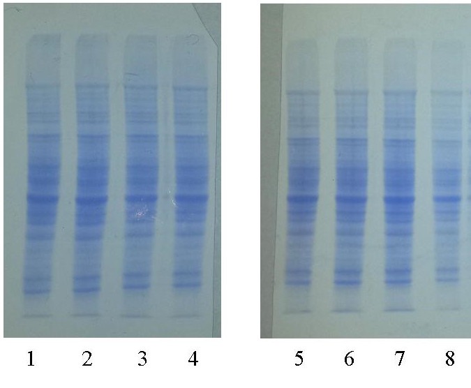
A PVDF blot of 8 identical lanes of mouse liver extract was divided in half, and lanes 5-8 were incubated in ProtoLift according to the standard protocol (see Figure 1). Both membranes were stained with Coomassie Blue to detect total protein levels. No detectable difference was seen between the control and the membrane incubated in ProtoLift Western Stripping Buffer.
PVDF Blots can be stripped and reprobed multiple times with ProtoLift
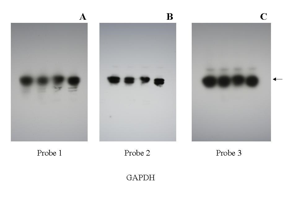
A PVDF blot of mouse liver extract was probed for GAPDH, then stripped and reprobed 4 times (see Figure 2). Panel A: first probing, Panel B: second probing, Panel C 4th probing. All 4 probings produced identical signals.
Additional information
Weight | 0.1 lbs |
---|
Protocol
Stripping Procedure
CAUTION: This procedure requires the use of mercaptoethanol, a hazardous substance. Work in a fume hood.
- Probe the membrane using your preferred protocol. (do not allow the membrane to dry before stripping).
- Rinse the membrane in two washes of PBS or TBS.
- Prepare a working ProtoLift Western Stripping Buffer solution by adding 2-mercaptoethanol to 0.7% (v/v). To prepare 10 ml of working solution, add 70μl mercaptoethanol to 10 ml ProtoLift stock solution.
- Place the membrane in a dish and add enough working solution to completely immerse the membrane. Alternatively the membrane can be sealed in a plastic bag with the working solution.
- Incubate at room temperature for 10 minutes with shaking. PVDF membranes will become transparent in the solution. This is normal and the membrane will return to its regular appearance after the stripping solution is removed.
- Wash the membrane three times for 10 minutes each with large volumes of PBS or TBS to remove the stripping solution and reducing agent. The blot is now ready to be reprobed.
Safety Overview
Safety Summary (see SDS for complete information before using product):
Appearance and Odor
Odorless, colorless solution.
Safety and Precautionary Overview
Warning! Irritant
Harmful if swallowed
Causes skin irritation.
Causes serious eye irritation.
Harmful if inhaled.
Wear protective gloves/protective clothing/eye protection/face protection.
If on skin, wash with plenty of soap and water.
If in eyes, rinse cautiously with water for several minutes. Remove contact lenses if present and easy to do. Continue rinsing.
Call a poison center or doctor/physician if you feel unwell.
- UV Shadowing
- Using PAGE to Determine Nucleic Acid Molecular Weight
- Uneven Staining
- The Polyacrylamide Matrix-Buffer Strength
- The Polyacrylamide Matrix
- The Mechanical and Electrical Dynamics of Gel Electrophoresis — Electrophoresis System Dynamics
- The Mechanical and Electrical Dynamics of Gel Electrophoresis – Ohm’s Law
- The Mechanical and Electrical Dynamics of Gel Electrophoresis – Intro and Sample Mobility
- The Electrophoresis Matrix
- The Agarose Matrix
- Staining Proteins Immobilized on Membranes
- Staining Protein Gels with Coomassie Blue
- SSCP Analysis
- Southern Blotting
- Smeared Bands
- Silver Staining Protein Gels
- Silver Staining DNA Gels
- Sanger Sequencing
- Sample Preparation for SDS-PAGE
- Sample Preparation for Native Protein Electrophoresis
- Sample Preparation for Native PAGE of DNA
- Sample Prep for Denaturing PAGE of DNA
- S1 Mapping
- Run Conditions in Denaturing PAGE
- RNA Mapping
- RNA Electrophoresis
- Ribonuclease Protection
- Restriction Digest Mapping
- Radioactive Emissions and the Use of Isotopes in Research
- Protein Fixation on Gels
- Primer Extension
- Preparing Denaturing DNA & RNA Gels
- Preparation of Denaturing Agarose Gels
- Preparation of Agarose Gels
- Pouring Sequencing Gels
- Post-Electrophoretic Visualization with Nuclistain
- PFGE and FIGE
- Peptide Mapping
- PCR Analysis: Yield and Kinetics
- PCR Analysis: An Examination
- Overview of Western Blotting
- Northern Blotting
- Native Protein Electrophoresis
- Native PAGE of DNA
- Multiphasic Buffer Systems
- Mobility Shift Assay
- Methylation & Uracil Interference Assays
- Method for Western Blotting
- Mechanism of Immunostaining
- Measuring Molecular Weight with SDS-PAGE
- Maxam & Gilbert Sequencing
- Manual Sequencing
- Isotachophoresis
- Isoelectric Focusing
- In Gel Enzyme Reactions
- Immunostaining with Alkaline Phosphatase
- Immuno-Electrophoresis / Immuno-Diffusion
- Horizontal and Vertical Gel Systems – Vertical Tube Gels
- Horizontal and Vertical Gel Systems – The Vertical Slab Gel System
- Horizontal and Vertical Gel Systems – The Horizontal Gel System
- Homogeneous Buffer Systems
- Heteroduplex Analysis
- Guide Strip Technique
- Gel Preparation for Native Protein Electrophoresis
- Gel Preparation for Native PAGE of DNA
- Gel Electrophoresis of RNA & Post Electrophoretic Analysis
- Gel Electrophoresis of PCR Products
- Faint bands, low background
- Faint Bands, High Background
- Ethidium Bromide Staining
- Enzyme Linked Immunosorbent Assay (ELISA)
- Electrophoresis Buffers-Choosing the Right Buffer
- Electrophoresis Buffers–The Henderson-Hasselbalch Equation
- DNase I Footprinting
- DNA/RNA Purification from PAGE Gels
- DNA/RNA Purification from Agarose Gels – Electroelution
- Differential Display
- Denaturing Protein Electrophoresis: SDS-PAGE
- Denaturing Polyacrylamide Gel Electrophoresis of DNA & RNA
- Coomassie Blue Stain- Troubleshooting
- Conformational Analysis
- Casting Gradient Gels
- Buffer Additives-Surfactants
- Buffer Additives-Reducing Agents
- Buffer Additives-Hydrogen Bonding Agents
- Blotches on Gel
- Biological Macromolecules: Nucleic Acids
- Biological Macromolecules – Proteins
- Autoradiography
- Autoradiographic Enhancement with Autofluor
- Automated Sequencers
- Analysis of DNA/Protein Interactions
- An Overview of Northern and Southern Blotting
- Alkaline Blotting
- Agarose Gel Electrophoresis of DNA and RNA – Uses and Variations
- Agarose Gel Electrophoresis of DNA and RNA – An Introduction
- Activity Stains